

What is Bacteriology?
- The field of microbiology that focuses on the study of bacteria is called bacteriology. In general, the scientific field of microbiology studies microscopic life forms such as bacteria, viruses, fungi, and other microbes. The study of morphology, physiology, genetics, and ecology of bacteria is known as bacteriology.
- Single-celled, prokaryotic bacteria can be found in a variety of places, including soil, water, and the human body. Bacteriologists do extensive research on the structure, development, metabolism, genetics, and functions of bacteria in a wide range of biological activities. Understanding the biology and behavior of bacteria, which can have both positive and negative effects on people, animals, and the environment.
- Bacteriologists also play a critical role in areas such as healthcare, agriculture, and environmental science, as they work to understand and control bacterial infections, develop antibiotics, study bacterial diseases, and look into the use of bacteria in biotechnology and bioremediation.
Importance of Bacteriology
Disease Diagnosis:
- Bacteriology is crucial for diagnosing infectious diseases caused by bacteria. Identifying the specific bacterial pathogen responsible for an illness is essential for prescribing the appropriate antibiotics or other treatments.
Public Health Surveillance:
- Bacteriology plays a key role in public health by monitoring and studying bacterial infections. Surveillance helps in understanding the prevalence, distribution, and trends of bacterial diseases, enabling the development of effective public health strategies.
Food and Water Safety:
- Bacteriology is instrumental in ensuring the safety of food and water. Bacterial contamination in food and water can lead to outbreaks of foodborne illnesses, and bacteriological testing helps identify and control these contaminants.
Industrial Applications:
- Bacteriology has industrial applications, including the production of antibiotics, enzymes, and other bio-based products. Understanding bacterial metabolism and genetics is essential for optimizing industrial processes and improving the efficiency of biotechnological applications.
Clinical Microbiology:
- Bacteriology is a key component of clinical microbiology laboratories, where diagnostic tests are performed to identify bacterial pathogens in patient samples. This information guides physicians in selecting appropriate treatments for bacterial infections.
Bacteria
- Bacteria are single-celled microorganisms that can be found in diverse habitats such as soil, water, and the human body. They are prokaryotes, meaning they lack a nucleus and other membrane-bound organelles, They are among the most abundant and diverse types of life on Earth, and they may be found in a wide range of habitats, including the interior of humans and the deepest parts of the ocean.
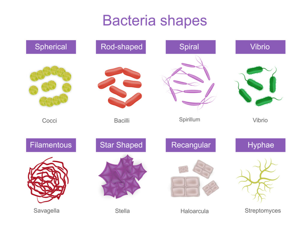
- Bacteria can have various shapes, including spherical (cocci), rod-shaped (bacilli), or spiral (spirilla). While some bacteria are harmful and can cause infections or diseases, others are beneficial and are involved in processes such as nutrient cycling, digestion, and the production of antibiotics.

Eukaryotic cell:
- Eu- true
- Karyote- nucleus
- The eukaryotic cell has a true membrane-bound nucleus, usually containing multiple chromosomes, a mitotic apparatus, a well-defined endoplasmic reticulum, and mitochondria.
Prokaryotic cell:
- Pro- primitive
- Karyote- nucleus
- The prokaryotic cell possesses naked DNA without associated basic proteins, divides mitotically by binary fission, and is bounded by a semi-rigid cell wall.

General properties of Bacterial Cell
- Typical prokaryotic cell.
- Contains both DNA and RNA.
- Most grow in artificial media.
- Replicate by binary fission.
- Almost all contain rigid cell walls.
- Sensitive to antimicrobial agents.
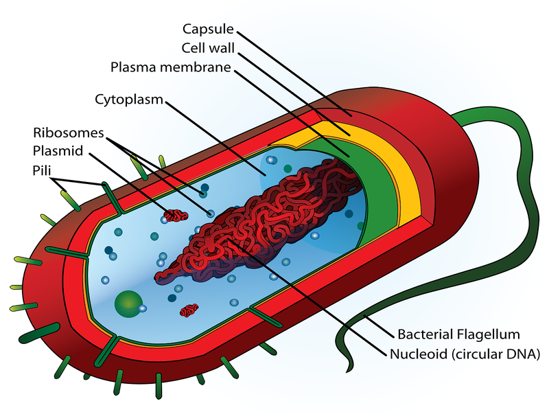
Structure of Bacteria
Bacterial structure is considered at three levels.
- Cell envelope proper: Cell wall and cell membrane.
- Cellular elements enclosed within the cell envelope: Mesosomes, ribosomes, nuclear apparatus, polyamines, and cytoplasmic granules.
- Cellular elements external to the cell envelope: Flagellum, Pilus, and Glycocalyx.
1. Cell envelope proper:
A.Cell wall
- Multi-layered structure and constitutes about 20% of the bacterial dry weight.
- The average thickness is 0.15-0.5 μm.
- Young and rapidly growing bacteria has thin cell wall but old and slowly dividing bacteria has thick cell wall.
- It is composed of N-acetyl Muramic acid and N-acetyl Glucosamine bones cross-linked with peptide chain and pentaglycine bridge.
Components of the cell wall of Gram-negative bacteria
- Peptidoglyca.
- Lipoprotein.
- Phospholipid
- Lipopolysaccharide
Components of the cell wall of Gram-positive bacteria:
- Peptidoglycan.
- Teichoic acid.
The function of the Cell wall:
- Provides shape to the bacterium.
- Gives rigidity to the organism’s Cell wall of Gram Positive & Gram Negative Bacteria.
- Protects from the environment.
- Provides staining characteristics to the bacterium.
- Contains receptor sites for phages/complements.
- Site of action of antibody and colicin.
- Contains toxic components to the host.
B.Cell membrane:
- It is also known as a cytoplasmic membrane (or) plasma membrane.
- It accounts for 30% of the dry weight of bacterial cells.
- It is composed of 60% protein, 20-30% lipids, and 10-20% carbohydrate.
Function of cell membrane:
- Regulates the transport of nutrients and waste products into and out of the cell.
- Synthesis of cell wall components.
- Assists DNA replication.
- Secrets proteins.
- Carries on the electron transport system.
- Captures energy in the form of ATP.
2. Cellular element enclosed within the cell envelope:
A. Mesosomes
- Mesosome is the extension of the plasma membrane which occurs in the bacterial cell.
- It is seen as an infolding of the plasma membrane and increases the surface area of activity for the cell.
- It is involved in DNA segregation during cell division and respiratory enzyme activity.
B.Ribosomes
- A tiny granule made up of RNA and proteins.
- They are the site of protein synthesis.
- They are free-floating structures that help in transferring the genetic code.
- It is composed of RNA(70%) and proteins(30%) and constitutes 90% of the RNA and 40% of the total protein.
- The ribosome monomer is 70s with two subunits, 30s and 50s.
C. Polyamines
- They are of three types
– Putrescin
-Spermidine
– Spermine - It is found in association with bacterial DNA, ribosomes, and cell membranes.
Function of polyamines:
- Antimutagenic.
- Prevent dissociation of 70s ribosome into subunits.
- Increase resistance of protoplast lysis.
D. Cytoplasmic granules: represent accumulated food reserves.
- Nature of granules:
– Glycogen
– Poly-beta hydroxybutyrate
– Babes-Ernst (Volutin)
E. Nuclear apparatus
- Well-defined nucleus and nuclear membrane, discrete chromosomes, and mitotic apparatus are not present in bacteria, hence the nuclear region of bacteria is named nuclear body, nuclear apparatus, and nucleoid.
- Bacterial genomes consist of single molecules of double-stranded DNA arranged in a circular form.
- In addition to the nuclear apparatus, bacteria may also contain extrachromosomal genetic material called plasmids.
- Plasmids play no role in the normal function of the bacterial cell but may provide some additional properties (e.g., virulence, drug resistance) that may facilitate the survival and spread of the microorganism.
3. Cellular elements external to the cell envelope
A. Glycocalyx (Capsule and slime layer)
- A capsule is a gel tightly attached to the cell envelope.
- Slime is a gel that can be easily washed from the cell membrane.
- All bacteria have at least a thin slime layer.
- The capsule is composed of polysaccharides and protein (D-glutamate of Bacillus anthracis).
Features of capsule:
- Usually weakly antigenic.
- Not necessary for feasibility.
- Provides virility.
- Prevents phagocytosis.
- Capsulated strains are always non-mobile.
- Visible by negative staining and capsule staining.
- Detected by Quelung phenomenon.
B. Flagella
- It is the organ of locomotion in bacterial cells and consists of three parts.
- These are. The filament. The hook. The basal body.
- The basal body and hook are embedded in the cell surface while the filament is free on the surface of the bacterial cell.
- Size: 3-20μm in length and 0.01-0.013μm in diameter.
- It is made of a protein called flagellin.
- The flagellar antigen in motile bacteria is named H (Hoch) antigen.
Their presence in bacterial cells is detected by
- Hanging drop preparation
- Motility media
- Special staining methods
- Silver impregnation methods
- Darkfield microscopy
- Electron microscopy
Flagellar Arrangement:
- Atrichous: Bacteria with no flagellum. e.g., Yersinia pestis.
- Monotrichous: Bacteria with single polar flagellum. e.g., Vibrio cholera.
- Lophotrichous: Bacteria with a bunch of flagella at one pole. e.g., Pseudomonas.
- Amphitrichous: Bacteria with a bunch of flagella at one pole. e.g., Pseudomonas.
- Peritrichous: Bacteria with flagella all over their surface. e.g., Salmonela species,E.coli.
Endoflagella (axial filament): It is an organ of mobility found in the periplasmic space of spirochetes.
C. Pili (fimbriae)
- It is a hair-like structure composed of protein (pilin).
Two types (Based on condition)
1. Common pili: The structure for adherence to the cell surface.
2. Sex pili: The structure for the transfer of genetic material from the donor to the recipient during the process of conjugation.
D. Spore
- A spore is a dormant, highly resistant, and non-reproductive structure that is formed in response to adverse environmental conditions.
- Spore formation is a survival strategy that allows some bacterial species to tolerate harsh conditions such as extreme temperatures, desiccation (drying), and exposure to chemicals or radiation.
- Resting cells that are capable of surviving adverse environmental conditions such as heat, drying, freezing, and action of toxic chemicals and radiation.
- Bacterial spores are smooth-walled and oval or spherical.
- It does not take up ordinary stains.
- It looks like areas of high refractivity under a light microscope.
- It is important in indicating the spread of disease and sterility of materials.
Spores are detected by
- Simple staining methods.
- Special staining methods.
Morphology of bacteria
When bacteria are visualized under a light microscope, the following morphology is seen.
- Cocci (singular coccus): Round or oval bacteria measuring about 0.5-1.0μmb in diameter. They are found in single, pairs, chains, or clusters.
- Bacilli (singular bacillus): Stick-like bacteria with rounded, tapered, square, or swollen ends; with a size measuring 1-10μm in length by 0.3-1.0μm in width.
- Coccobacilli (singular coccobacillus): Short rods.
- Spiral: Spiral-shaped bacteria with regular or irregular distance between twisting.
Eg. Spirilla and Spirochaetes.
References
- https://www.cartercenter.org/resources/pdfs/health/ephti/library/lecture_notes/env_occupational_health_students/medicalbacteriology.pdf
- https://www.nature.com/subjects/bacteriology
- https://www.sciencedirect.com/topics/immunology-and-microbiology/bacteriology
- https://www.ncbi.nlm.nih.gov/books/NBK8120/
- https://microbenotes.com/category/bacteriology/
- https://repository.poltekkes-kaltim.ac.id/1153/1/medical%20microbiology.pdf
- https://sc.uobaghdad.edu.iq/wp-content/uploads/sites/64/mainfiles/lectures/bio/%D8%A7%D9%84%D8%A8%D9%83%D8%AA%D8%B1%D9%8A%D8%A7.pdf
- https://apsjournals.apsnet.org/series/bacteriology
- https://www.lecturio.com/concepts/bacteriology-overview/
- https://vetmed.iastate.edu/vdl/laboratory/bacteriology
- https://dhhr.wv.gov/ols/labs/Pages/Bacteriology.aspx