
What is Gram Staining?
Table of Contents
hide
- Developed by the Danish Bacteriologist Hans Christian Gram in 1884. Most bacteria are differentiated by their gram reaction due to differences in their cell wall structure.
- Gram staining is a differential bacterial staining method used to distinguish between Gram-positive and Gram-negative bacteria based on their cell wall composition.
- It is the most significant and extensively utilized staining method in bacteriology, particularly in medical bacteriology.
- Gram-positive bacteria are bacteria that stain purple with crystal violet after decolorizing with acetone-alcohol.
- Gram-negative bacteria are bacteria that stain pink with the counter stain (safranin) after losing the primary stain (crystal violet) when treated with acetone-alcohol.
- Gram-positive bacteria have thick, dense, largely non-porous walls. While Gram-negative bacteria have thin walls surrounded by lipid-rich membranes.
Objectives of Gram Staining
- To distinguish between Gram-negative and Gram-positive bacteria.
- To investigate the morphology of bacteria.
Requirements of Gram Staining
- Sample bacterial colonies or suspension
- Gram Staining Kit (Reagents)
- Glass slide
- Inoculating loop
- Bunsen burner
- Staining rack
- Stop watch
- Wash bottle (or Tap water)
- Microscope with 100X objective lens (compound microscope)
Gram Stain Reagents
The following types of chemicals and dyes are used in the Gram staining process:
1. Primary Stain (Crystal Violet)
- It is used in ball pens, detergents, fertilizers, and fibres as well as textiles, papers, and other materials.
- It is utilized in molecular biology and microbiology for staining DNA, histology slides, and bacteria, among other things.
- It is utilized in sterilization and disinfection since it possesses antibacterial and antifungal qualities as well.
- In Gram Staining, It provides a violet colour to Gram-positive bacteria.
Crystal Violet Preparation
- In 20 ml of 95% ethyl alcohol, dissolve 2 g of crystal violet.
- Dissolve 0.8 g ammonium oxalate monohydrate in 80 ml deionized water.
- Mix the crystal violet and ammonium oxalate monohydrate solutions to make the crystal violet stain.
2. Mordant (Gram’s Iodine solution)
- Gram staining uses this aqueous solution of potassium iodide and iodine as a mordant.
Gram’s Iodine Preparation
- In 300 ml of distilled water, dissolve 1 gram of iodine and 2 grams of potassium iodide.
- After mixing properly until the iodine dissolves, keep the mixture in a dark bottle.
3. Decolourizer (Acetone)
- It is either 95% ethanol or acetone, or a 1:1 volume ratio of both substances together.
- A mixture of acetone-alcohol is recommended because it decolorizes more quickly than ethanol.
Acetone-alcohol Preparation
To make 1 litre:
- Acetone……….500 ml
- Absolute ethanol or methanol……….475 ml
- Distilled water………. 25 ml
4. Counter Stain (Safranin)
- Common counter stains used in the Gram staining process are safranin and diluted carbol-fuchsin.
- It is a basic dye that reacts with the negatively charged membrane and cell wall components.
Safranin Preparation
- Mix 2.5 grams of safranin-O with 100 ml of 95% ethanol.
- To prepare a working solution, Mix 90 ml of distilled water with 10 ml of the above solution.
Dilute carbol-fuchsin Preparation
- In 10.0 ml of 95% ethyl alcohol, dissolve 0.3 grams of basic fuchsin.
- To 95.0 ml of distilled water, add 5.0 ml of melted phenol crystals.
- Add the 5% phenol solution to the fuchsin solution and leave it overnight.
Importance of fixing the smears
Three goals are achieved through fixation:
- It kills the organisms.
- It causes the organisms to stick to the slide.
- It changes the organisms to make them more receptive to stains (dyes).
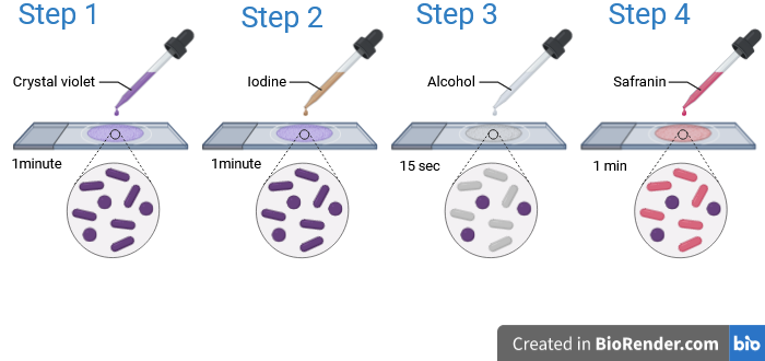
Procedure of Gram Staining
- Take a clean dry glass slide and label it.
- Sterilize the inoculating loop over flame.
- Prepare a thin uniform smear from the culture or the specimen.
- Dry the smear in air.
- Fix the smear over flame.
- Cover the smear with crystal violet solution for 1 minute. wash with tap water.
- Cover the smear with gram’s iodine solution for 1 minute. wash with tap water.
- Add decolorizer acetone-alcohol for 15 seconds. wash with tap water.
- Cover the smear with safranin for 1 minute. wash with tap water.
- Dry the smear in air.
- Examine the smear with an oil immersion objective to look for bacteria.
Result of Gram Staining
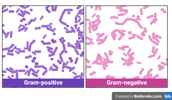
Gram-positive bacteria………. violet or Purple.
Gram-negative bacteria……….pink or red.
Examples of Gram-positive bacteria
- Gram-positive cocci (spherical): Streptococcus spp, Staphylococcus spp, Enterococcus spp., ect.
- Gram-positive bacilli (cylindrical): Clostridium spp., Lactobacillus spp., Corynebacterium diphtheria., etc.
Examples of Gram-Negative bacteria:
- Gram-negative cocci (spherical): Neisseria meningitis, Neisseria gonorrhoeae (STD).
- Gram-negative bacilli (cylindrical): E. coli, Salmonella spp., Shigella spp., Pseudomonas spp., Klebsiella spp.
Applications of Gram Staining
- Used in studies to divide bacteria into Gram-positive and Gram-negative groups.
- Used in diagnostic laboratories to find out the pathogen’s identification.
- Used in hospitals to select an antibiotic range before fully identifying the organism to be treated.
- Used in the study of bacterial morphology.
Limitations of Gram Staining
- Not able to stain Mycobacterium spp (Acid Fast Bacilli) or bacteria lacking a cell wall, such as Mycoplasma spp.
- Require more than one reagent.
- False gram-positive results might be identified by under-decolourization, whereas false gram-negative results could be identified by over-decolourization.
- Using the Gram stain, not all bacteria are visible.
- It might be difficult to identify the correct Gram stain reactions in smears that are very thick or viscous because they retain an excessive amount of primary stain. It’s possible that gram-negative bacteria may not decolourize properly.
- For mycobacteria and spirochetes, the Gram staining method is not advised.
Read Also
References
- Gram Staining: Principle, Procedure, Results-Microbe Online
- Gram Staining- Principle, Reagents, Procedure, Steps, Results- Microbe Notes
- Preparation of Gram stain Reagents-Microbe Online
- Gram Stain, What is a Gram stain? Cleveland Clinic
- Gram Stain: MedlinePlus
- Gram Stain: Testing.com
- Gram Staining:Principle, Procedure, Interpretation, Examples and Animation-Laboratoryinfo.com
- Gram Stain: Purpose, Theory & Procedure-Study.com
- Gram Staining: Principle, Procedure and Results-microbiologie-clinique.com
2 thoughts on “Gram Staining: Definition, Reagents, Procedure, Results”